Medically reviewed by Dr. Hyun Joon Lee, MD. Board-certified in Family Medicine, Integrative Holistic Medicine, and Obesity Medicine. Founder and Medical Director, SeeBeyond Medicine.
What deep TMS and rTMS have in common
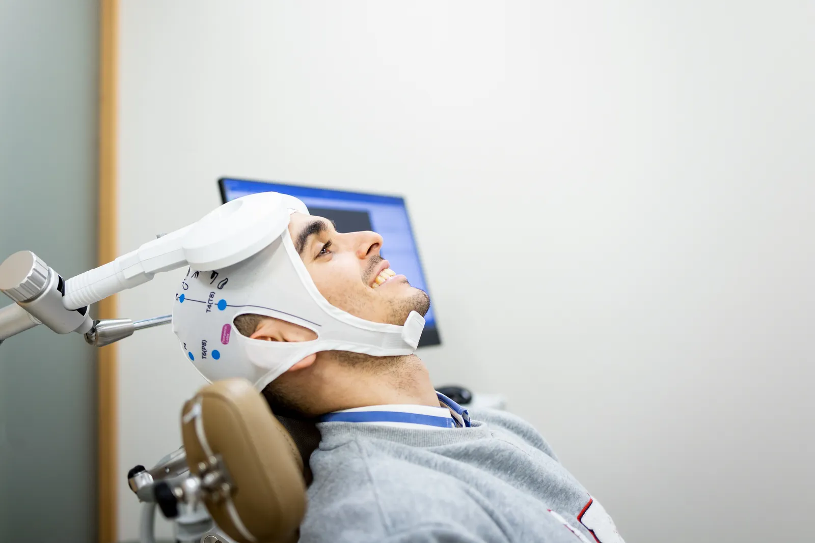
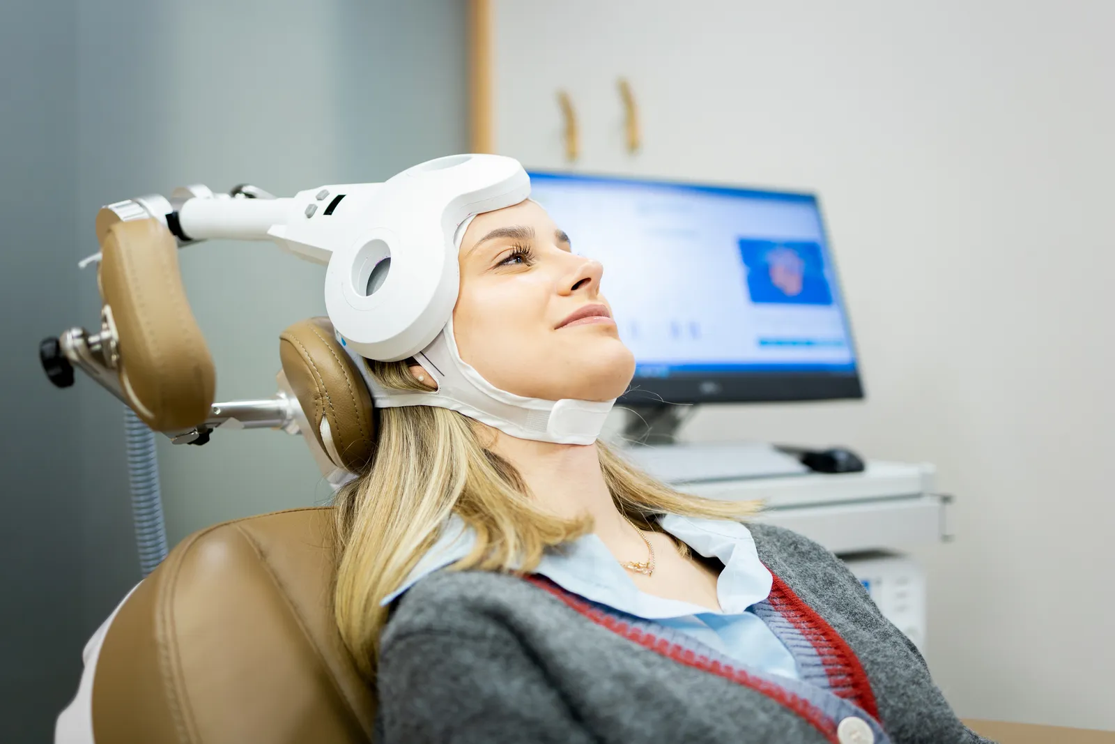
Transcranial magnetic stimulation uses a coil placed against the scalp to generate brief magnetic pulses. Those pulses induce an electric field in nearby brain tissue. When the field is strong enough and correctly oriented, it depolarizes axons and activates neurons in the targeted structure (FDA De Novo summary DEN170078).
Repetitive TMS, shortened to rTMS, means those pulses arrive in timed trains rather than one at a time. Deep TMS is a form of rTMS.
The distinction is therefore not pulses against no pulses. It is the shape of the coil producing the field, and the shape of the field that reaches your brain.
Figure-8 coils produce a narrow, shallow field
A figure-8 coil holds two circular windings side by side. The currents run in opposite directions, so the induced fields add together at the crossover point and cancel elsewhere. That geometry creates a tight hot spot on the cortical surface.
Precision is the payoff. In a simulation study of 50 coil designs, coils producing figure-8 field patterns reached a tangential spread as low as 5 cm², against 34 cm² for coils producing circular field patterns (Deng, Lisanby and Peterchev, Brain Stimulation, 2013).
The cost is reach. The induced field falls off quickly with tissue depth, which is why figure-8 protocols for depression target a surface structure, the left dorsolateral prefrontal cortex.
H-coils spread the field wider and deeper
The H-coil, named the Hesed coil, was designed by Roth, Zangen and Hallett in 2002 for exactly this limitation. Its windings are dispersed tangentially along the skull rather than concentrated at one crossover point, which slows the rate at which the field decays.
Physiological testing supported the design. Comparing coils against motor thresholds in healthy volunteers, the rate of decrease in stimulation intensity as a function of distance was markedly slower for the H-coil than for a standard figure-8 coil (Zangen and colleagues, Clinical Neurophysiology, 2005).
Modelling work points the same direction. A finite element comparison found deeper penetration for the H1 coil than the figure-8, a difference of roughly 0.7 cm in half-value depth, alongside wider field distribution and more gradual decay (PLOS ONE, 2017).
Depth is a trade-off rather than a straight upgrade
Every one of those 50 simulated coils obeyed the same rule. Coils with greater half-value depth could not also be focal, and the authors concluded that deep-brain designs are unfocal and may carry increased risk of seizures and other side effects.
That finding matters more than any marketing comparison. Reaching a deeper structure means stimulating a larger volume of tissue on the way there.
So the useful question is not which coil is stronger. It is which target your diagnosis points to, and how much surrounding cortex you are willing to recruit to reach it.
Side-by-side comparison
| Feature | Figure-8 coil rTMS | H-coil deep TMS |
|---|---|---|
| Coil geometry | Two adjacent windings, fields summing at one crossover point | Windings dispersed tangentially along the skull |
| Field depth | Shallower half-value depth, with rapid decay through tissue | Roughly 0.7 cm greater half-value depth than figure-8 in modelling |
| Focality | More focal, tangential spread as low as 5 cm² in simulation | Broader spread, so deeper reach recruits more cortex |
| Typical session length | 37.5 minutes for the standard 10 Hz protocol, about 3 minutes for iTBS | 20 minutes of stimulation in the pivotal depression protocol, 18 minutes in the cleared OCD protocol |
| Example cleared indications | Adult MDD, OCD as an adjunct, anxiety symptoms in adult MDD, adolescent MDD as an adjunct | MDD, OCD, short-term smoking cessation in adults |
What each device is cleared to treat
For most patients this is the difference that decides which room they sit in. Clearance is granted per coil and per indication, not to the category as a whole.
On the figure-8 side, NeuroStar was first cleared for adults with major depressive disorder in 2008. Its adolescent indication, an adjunct for patients aged 15 to 21, was cleared in 2024 (FDA 510(k) K231926).
On the H-coil side, the H7 coil received De Novo clearance for OCD in August 2018. That protocol targets the dorsomedial prefrontal cortex and anterior cingulate cortex at 20 Hz for 18 minutes.
A separate H4 coil was cleared in 2020 as an aid to short-term smoking cessation in adults. It followed a multicenter double-blind randomized controlled trial in 262 chronic smokers that reported a significantly higher four-week continuous quit rate with active stimulation (Zangen and colleagues, World Psychiatry, 2021).
Session length is a separate variable
Chair time is often confused with coil type, and the two move independently. The pivotal H1 depression protocol delivered 18 Hz stimulation at 120% of resting motor threshold, with 20 minutes of stimulation inside a 30-minute session (Levkovitz and colleagues, World Psychiatry, 2015).
Figure-8 protocols have moved in the other direction. The THREE-D trial found intermittent theta burst stimulation, delivered in about 3 minutes, non-inferior to the standard 37.5-minute 10 Hz protocol in treatment-resistant depression (Blumberger and colleagues, The Lancet, 2018).
How the choice gets made in practice
Diagnosis narrows the field first. An OCD presentation, a depression presentation, and a tobacco use disorder presentation do not lead to the same coil.
History narrows it further. Prior medication trials, seizure risk, implanted metal, current medications, and sleep patterns all belong in that conversation before a protocol is selected.
Device availability and coverage then shape what is practical. SeeBeyond Medicine runs a deep TMS program covering more than 30 conditions, with a 91% response rate across the program, and candidacy is still assessed case by case.
Key Takeaways
- Both approaches deliver repetitive magnetic pulses; the coil geometry is what changes the depth and breadth of the field.
- Figure-8 coils focus a shallow, narrow field, while H-coils spread a broader field to deeper structures.
- Depth is a trade-off rather than a straight upgrade, because broader stimulation is less focal.
- The clearest practical difference for patients is which conditions each device is cleared to treat.
Related Resources
Start with our complete guide to deep TMS therapy for the full cluster, including candidacy, protocol structure, and side effects.
For condition-specific detail, read about deep TMS for anxiety and mood disorders, deep TMS for OCD, deep TMS for PTSD, and deep TMS for insomnia.
Patients weighing several neuromodulation and infusion options often compare TMS against ketamine therapy during the same consultation.
Coil geometry is a genuine clinical variable, and it is one of several. The rest of the decision depends on your diagnosis, your treatment history, and what your testing has already shown.
If you want that assessment done properly, deep TMS therapy at SeeBeyond Medicine is delivered under physician supervision at our Scarsdale, NY office in Westchester County and our Greenwich, CT office in Fairfield County. You can schedule a consultation to review whether deep TMS suits your situation.
This article is for general education and does not replace a medical evaluation. Treatment decisions depend on your history, medications, and lab work. Speak with a qualified clinician before starting, stopping, or changing any therapy.